
The Story Behind our Sample Prep Automation Innovations
.jpg)
Our mission is to democratize spatial biology discovery and biomarker capabilities through advanced technologies that increase accessibility, efficiency, and data quality of analytical techniques.
Parhelia Bio emerged from a shared vision at Stanford University.

As pioneers in the Spatial Biology field since its inception, we are uniquely positioned to lower the bar for anyone inspired to adopt these technologies in their research.
Co-founders Nikolay Samusik and Yury Goltsev were postdocs in the lab of Professor Garry Nolan at Stanford, where they co-developed the CODEX ultra-multiplexed spatial biology technology. They co-founded Akoya Biosciences (NASDAQ: AKYA) to commercialize CODEX (now PhenoCycler) and later acquired Opal (from Perkin Elmer) to offer critical technologies in spatial biology.
As front-line scientists pushing the boundaries of Spatial Biology, Nikolay, Yury, and Garry recognized the limitations of manual processing in preparing and staining samples for imaging analysis. Together, they founded Parhelia Biosciences to tackle these challenges head-on and enable researchers and clinicians to translate spatial biology discoveries into meaningful, biomarker-based outcome benefits for patients through reliable, consistent, and affordable automated solutions.
Purpose-Built Automation for the Spatial Biology Community
The speed and breadth of spatial biology's innovation have only increased the need for universal and high-throughput solutions to pre-imaging sample preparation.
We’re engineering consistent, reagent-saving, hands-free automated solutions to sample prep and staining bottlenecks so you can image
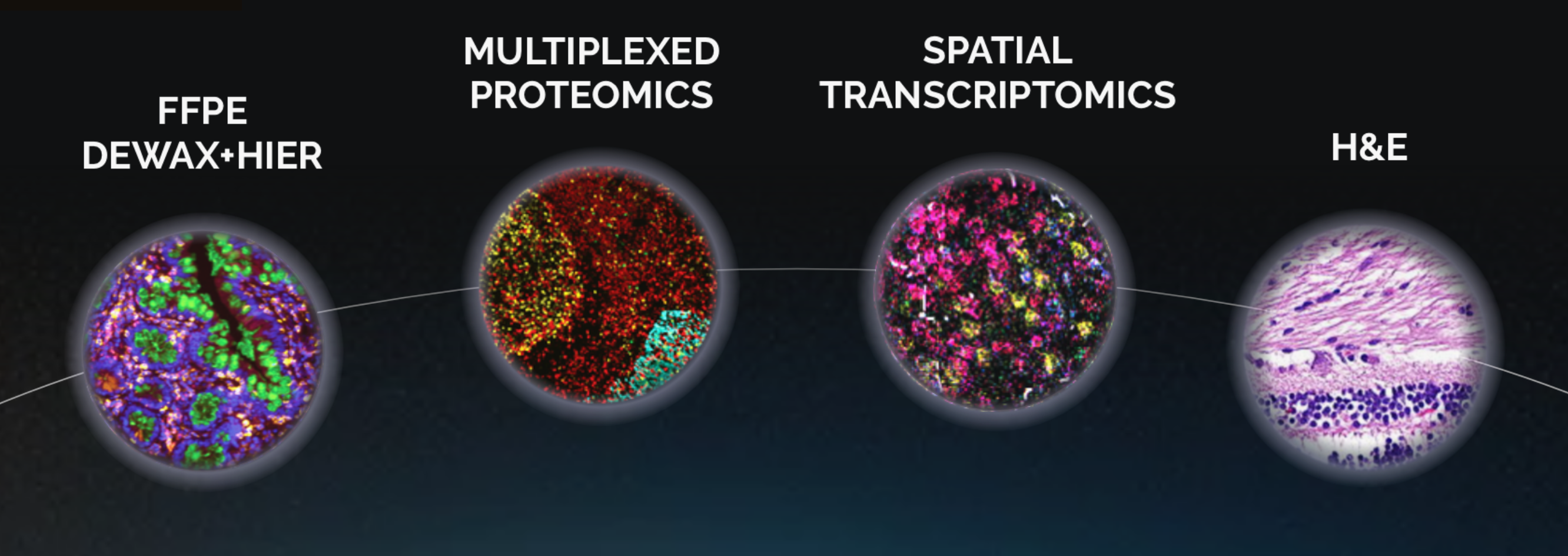
From basic stains to advanced protocols like PhenoCycler, GeoMx and Xenium, we simplify lab workflow and make tissue auto-staining accessible to all researchers on a budget.
Join us in advancing spatial biology with cutting-edge technology tailored to your needs.more with less.
The Team behind the scenes
Nikolay Samusik

Katherine Billet

Nels Wedin

Kevin Euell

Mike Stadnisky

Michael Craft
Mary Chandler Grisillo

Yury Goltsev

Prof. Garry Nolan
The timeline of Parhelia

2021
A small set of PAR2 instruments were distributed to Early Adopter labs for assay development and verification. Additional staining methods and automation protocols were developed for Parhelia devices (H&E, IHC, multiplexed IF, Opal, RNAscope FISH).
2022
Based on the Early Adopter feedback, the Omni-Stainer™ was designed and released. Parhelia enters an official partnership with Opentrons for a complete low-cost auto-staining solution.
2023
Based on the Early Adopter feedback, the Omni-Stainer™ was designed and released. Parhelia enters an official partnership with Opentrons for a complete low-cost auto-staining solution.
2024 - May
Parhelia Biosciences Announces the Launch of the Parhelia Spatial Station™ – A Revolutionary Sample Prep System for Spatial Biology
2024 - June
Testimonials
Staff Scientist
nih
• Dr. Noemi Kedei
senior scientist
Washington University in st. louis
• Dr. Ruan Medrano
Partners


Hamilton
Parhelia Bio is pleased to be partnering with Hamilton to bring the next generation of sample automation for spatial biology and flow cytometry with the Parhelia Spatial Station™.
Opentrons
Parhelia Bio is pleased to be partnering with Opentrons to offer competitive pricing on a complete tissue auto-staining solution. Omni-Stainer™ is compatible with most liquid handling robots, but if you don’t have one, we recommend our partner Opentrons OT-2.
.webp)
